Below-knee (transtibial) amputation
Below-knee amputation preserves the knee. Aim for a healed, comfortable residuum with viable padding, useful knee movement and tension-free coverage.
Written September 12, 2026 · Mr Razhan K Ali
PURPOSE & INDICATIONS
Below-knee amputation (BKA) divides the tibia and fibula while preserving the knee. Aim for a healed, comfortable, well-padded residuum with useful knee movement. Knee preservation benefits rehabilitation only when the residual limb can heal and function.[1–3]
Indications include irreversible ischaemia, uncontrolled infection or tissue destruction, and a painful non-functional limb when further salvage is inappropriate. For chronic limb-threatening ischaemia (CLTI), NICE advises against major amputation until a vascular multidisciplinary team has considered all revascularisation options. Revascularisation may still be worthwhile to enable a BKA even when the foot cannot be saved. Urgent source control takes priority when infection threatens life.[1,4,5]
Consent
Explain why amputation is proposed, why this level is appropriate, and the alternatives: further salvage, another amputation level, or non-operative/palliative care. Discuss bleeding/transfusion, infection, delayed healing or wound breakdown, revision or conversion to a higher level, residual-limb and phantom pain, neuroma, contracture, thromboembolism, cardiopulmonary complications and death. Establish realistic expectations for transfers, wheelchair use and possible prosthetic walking; neither pain relief nor prosthetic use is guaranteed.[1–3]
ANATOMY & PLANNING
Know the compartments
| Compartment | Structures that matter during BKA |
|---|---|
| Anterior | Anterior tibial vessels and deep peroneal nerve within the dorsiflexor compartment. |
| Lateral | Peroneus longus/brevis and superficial peroneal nerve. |
| Superficial posterior | Gastrocnemius and soleus: the principal source of posterior muscle padding. |
| Deep posterior | Posterior tibial and peroneal vessels, and tibial nerve. The peroneal vessels lie close to the fibula. |
The three principal arterial pedicles are therefore anterior tibial, posterior tibial and peroneal. Do not assume that an intact tibioperoneal trunk will be encountered at every amputation level. Preserve the perforators and tissue attachments supplying the chosen flap.[1,6]
Assess perfusion at the proposed level, infection extent, scars, previous bypasses, knee movement, baseline mobility, frailty and rehabilitation goals. Doppler assessment and selective perfusion testing supplement clinical judgement; no single pulse, pressure or transcutaneous oxygen threshold guarantees healing. Check imaging for tibial metalwork before committing to the osteotomy.[1–3,7]
Operative strategy
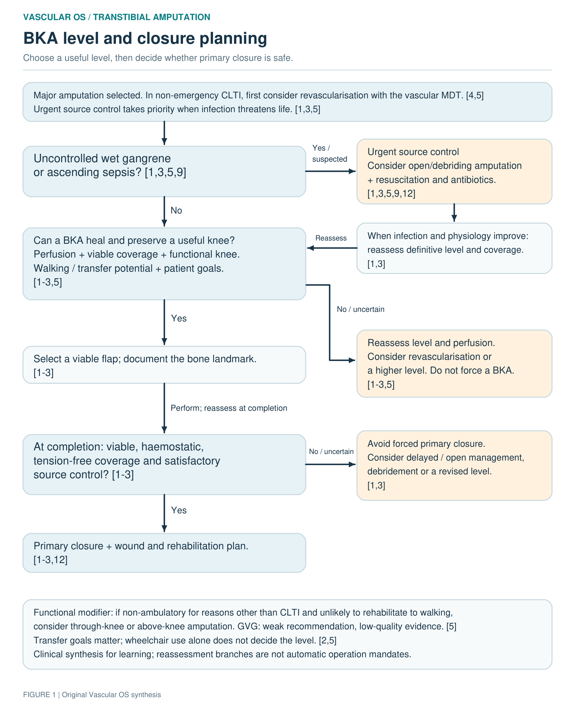
Assess walking and transfer potential. Preserve a functional knee when healing and useful transfers or prosthetic use are realistic. If non-ambulatory for reasons other than CLTI and unlikely to regain useful mobility, consider through-knee or above-knee amputation to avoid a non-healing, contracted BKA residuum. Do not equate temporary ischaemic immobility or wheelchair use alone with absent rehabilitation potential.[2,3,5,17]
Fixed knee flexion, severe knee arthropathy or a very short residual tibia may favour a higher level. Assess functional knee position with rehabilitation and prosthetic input; published contracture thresholds differ and should not become a universal cut-off.[1,2,5]
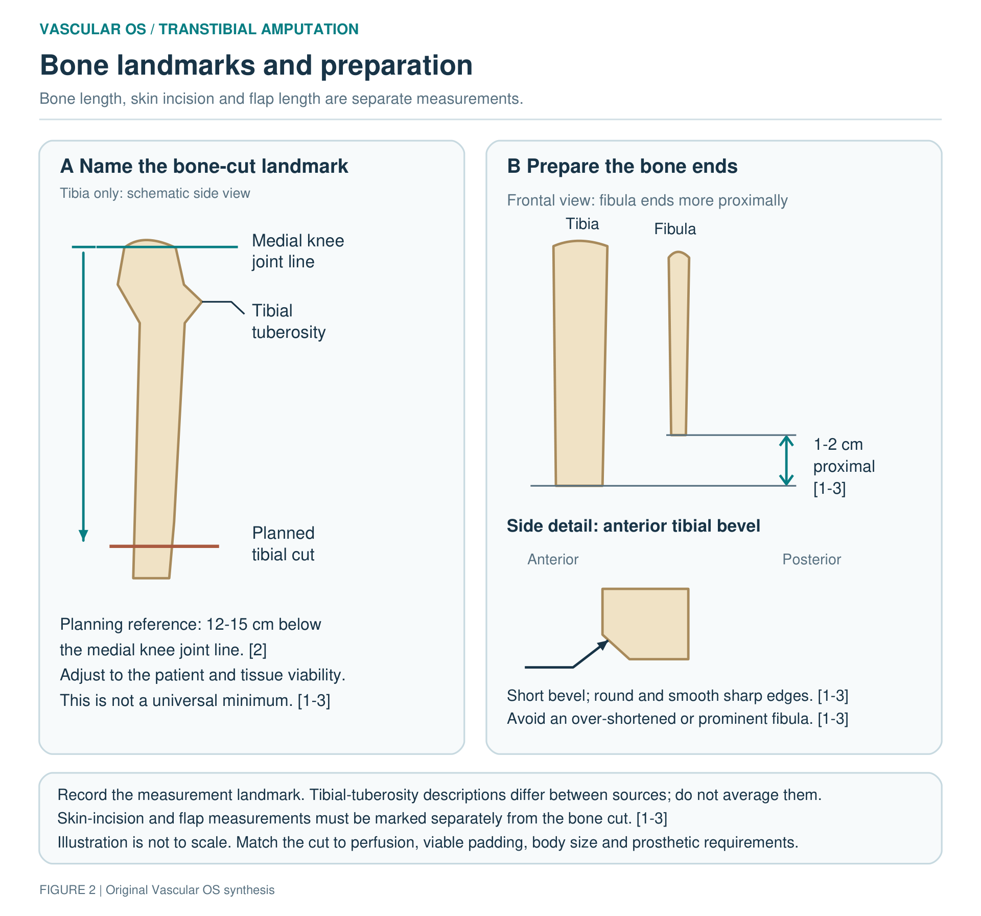
Plan the bone cut from a named landmark. One planning reference is 12–15 cm below the medial knee joint line, not a universal minimum.[2] Adjust the cut for body size, perfusion, viable coverage, patellar-tendon preservation and the prosthetic plan. Tibial-tuberosity measurements differ between operative descriptions; skin-incision and flap dimensions are separate again. Do not convert or average these measurements.[1–3]
Choose the flap before cutting. The Burgess long-posterior flap uses posterior calf tissue to cover the bone ends. The skew technique uses separate anteromedial and posterolateral fasciocutaneous flaps over posterior gastrocnemius coverage. Scars, tissue viability and the reconstructive plan determine suitability. Randomised evidence has not established superiority of either design.[1–3,8,9]
Choose primary closure or staging. Uncontrolled wet gangrene or ascending sepsis may require an initial open/debriding amputation, followed by definitive BKA when infection and physiology are controlled. Uncertain flap viability may justify delayed closure. Do not close contaminated or non-viable tissue simply to complete a one-stage procedure.[1,3,9]
Decision diagram — Level and closure planning
SETUP
For planned UK major-amputation surgery, a consultant vascular surgeon operates or is present to supervise a senior trainee on a daytime list. Urgent source control must not wait for an elective list.[12]
Confirm side, level, flap design, consent and the contingency for a higher or staged amputation. Position supine, with the knee accessible and the limb supported; isolate the diseased foot and protect the contralateral heel. Have vascular clamps and ligatures, an amputation knife, periosteal elevator, bone guards, saw and rasp available; add a drill if myodesis is planned.[1–3,7]
Agree anaesthesia and multimodal analgesia before surgery. The 2026 UK Delphi consensus favours an ultrasound-guided proximal sciatic perineural catheter at or above the popliteal fossa for BKA. A directly placed tibial catheter may be a practical alternative but provide incomplete coverage. Greater reliability of ultrasound-guided versus surgical placement did not reach consensus. Follow the agreed anaesthetic/acute-pain plan for supplementation, monitoring and removal.[10]
Tourniquet use is a case-specific choice. A 2024 review of major amputations—one randomised trial and six cohort studies—found lower intra-operative blood loss, a smaller postoperative haemoglobin fall and shorter operating time. It found no significant difference in total blood loss over the first 3–4 postoperative days, transfusion, surgical-site infection, revision or mortality; certainty was low or very low for every outcome.[11] If used, protect the skin and release the tourniquet before final haemostasis and flap assessment.[3,7]
Give peri-incision antibiotics and document the plan for established infection. Review haemoglobin, renal function, glucose, nutrition, antithrombotics and postoperative level of care. Involve rehabilitation services before non-emergency surgery where feasible.[1,5,12]
PERFORM
Long-posterior-flap technique
Phase A — Mark the reconstruction and create viable flaps
Mark the knee joint line, tibial tuberosity, intended bone cut and skin incisions separately. For a Burgess design, the conventional rule of thirds uses an anterior transverse incision spanning approximately two-thirds of the calf circumference and a posterior flap extending distally by approximately one-third of that circumference. Tailor the design to tissue thickness, scars and viability rather than treating the ratio as a rigid template.[1,3,7]
Incise skin, subcutaneous tissue and fascia along the markings. Control superficial veins as encountered. Avoid bevelled skin cuts and unnecessary undermining; maintain the planned posterior flap's tissue attachments.[1–3]
Checkpoint: viable flap edges and enough posterior tissue to cover the planned bone ends without tension.[1–3]
Phase B — Divide the anterior and lateral compartments under control
Divide the anterior and lateral muscle groups progressively. Identify and secure the anterior tibial artery and accompanying veins, keeping the deep peroneal nerve separate. Treat other muscular vessels as encountered rather than relying on a blind, en-masse clamp across the compartment.[1,2,6]
Identify any previous bypass before division. Prior prosthetic grafts increase stump-infection risk, and complete removal of synthetic material can reduce that risk.[1] Plan removal of infected or exposed material as part of source control. The extent of removal of uninvolved proximal graft is an individual operative judgement, not a universal requirement derived from these data.[1,3]
Checkpoint: the anterior pedicle is controlled, the bone is accessible, and the posterior flap has not been inadvertently divided or devascularised.[1–3]
Phase C — Make protected bone cuts and remove pressure points
Expose only enough tibia and fibula for safe division; avoid extensive periosteal stripping. Protect the posterior structures with a bone guard or retractor (or big gauze-swab). Divide the tibia at the planned level and the fibula approximately 1–2 cm proximal to the tibial cut. The order of division varies; protection of the soft tissues and the relative bone lengths are the important endpoints.[1–3,7]
Create a short bevel on the anterior tibial edge, round sharp corners and smooth both bone ends with a rasp. Remove bone debris. Do not leave a prominent fibular end, but do not shorten the fibula excessively: that can produce an unfavourable conical residuum.[1–3]
Checkpoint: smooth, protected bone ends; a shorter fibula; sufficient viable soft tissue for coverage.[1–3]
Phase D — Complete the posterior division and manage nerves
With the bone ends controlled, divide the remaining tissues along the posterior surfaces of the tibia and fibula to develop the long posterior flap. Preserve the gastrocnemius and its fascial attachments. Identify and secure the posterior tibial and peroneal vascular pedicles as they are exposed, separating the tibial nerve from the vessels. Complete haemostasis after removal of the limb.[1–3,6]
Identify the major nerves encountered, including the tibial, deep and superficial peroneal, sural and saphenous nerves. Conventional management is sharp division under gentle tension so the nerve ends retract into healthy proximal soft tissue, away from the scar and pressure points. Avoid unnecessary crushing and forceful traction. Evidence that traction neurectomy changes acute or chronic pain outcomes is scarce and equivocal; it does not guarantee prevention of neuroma or phantom pain.[1–3,10]
Checkpoint: all major pedicles are accounted for, nerve ends are protected, and the retained posterior muscle is viable.[1–3]
Phase E — Tailor the muscle envelope and secure coverage
Bring the posterior flap forwards and assess the coverage before trimming. Preserve enough viable gastrocnemius and fascia to cushion the tibial end. Trim or remove soleus when it is non-viable or creates excessive bulk; routine wholesale excision is not required.[1–3]
Secure posterior fascia/muscle coverage to the anterior fascia and periosteum without tension. Myodesis anchors gastrocnemius to the anterior tibia through drill holes; myoplasty joins muscle groups. Formal myodesis is an option, not an established requirement for every dysvascular BKA.[1,2]
Checkpoint: stable padding over the tibia without exposed bone, redundant muscle bulging through the wound or tight closure.[1–3]
Phase F — Recheck haemostasis, close and protect the knee
Release any tourniquet. Inspect each pedicle, the muscle surfaces, bone ends and flap perfusion. Irrigate away residual debris. If viability or source control remains doubtful, reassess the level or staged plan rather than forcing primary closure.[1–3]
Approximate the deep fascia and close the skin without strangulation. Avoid sacrificing viable corners merely to eliminate dog-ears.[1–3] Although operative descriptions commonly include a drain, the 2024 ERAS/SVS consensus suggests against routine drainage on low-certainty evidence. Use a drain only for a specific indication; it does not replace haemostasis.[1,2,17] Apply a protective, non-constricting dressing; pad any splint or immobiliser and check for pressure over the patella and other bony prominences.[1–3]
Checkpoint: haemostatic, viable, tension-free coverage with a clear wound-inspection and knee-positioning plan.[1–3]
Skew flap: what changes
A skew BKA is not simply a Burgess incision rotated obliquely. Create anteromedial and posterolateral fasciocutaneous flaps with their vascular attachments preserved. The posterior gastrocnemius muscle flap is brought forwards separately and anchored over the tibia; the skin flaps then meet obliquely, away from the cut bone. The principles of pedicle control, bone preparation, nerve management and tension-free closure remain unchanged.[1,2]

TECHNICAL PEARLS & PITFALLS
Separate bone length from flap length. A familiar centimetre value measured from the wrong landmark can compromise the reconstruction.[1–3]
Balance padding and bulk. Tight closure over bulky muscle risks ischaemia; excessive trimming leaves unprotected bone.[1–3,7]
Protect knee extension without pressure injury. Check the patella beneath any immobiliser.[1–3]
COMPLETION
Document level and measurement landmark, flap type, tissue viability, vessel and nerve management, previous graft material, muscle fixation, drains/catheters and any planned return to theatre. Specify antibiotics, thromboprophylaxis, analgesia, wound review, knee positioning and rehabilitation. Complete National Vascular Registry documentation.[1–3,7,12]
COMPLICATIONS & RESCUE
| Problem | Recognition and response |
|---|---|
| Bleeding/haematoma | Increasing swelling, pain, wound bleeding or physiological deterioration require urgent assessment and resuscitation. Ongoing bleeding or a collection threatening flap viability may need operative evacuation and haemostasis—not tighter bandaging.[1,3] |
| Flap ischaemia, dehiscence or infection | New pallor, coolness, blistering, progressive necrosis, drainage or systemic illness require wound inspection. Remove external constriction, reassess perfusion and source control, and consider debridement, open management or revision to a higher level.[1,3] |
| Knee flexion contracture | Prevent prolonged flexed positioning; maintain extension and begin appropriate range-of-motion exercises. Escalate loss of extension early to rehabilitation services rather than waiting until prosthetic fitting.[1,2] |
| Persistent residual-limb pain | Exclude ischaemia, infection, haematoma, prominent bone and socket pressure before attributing pain to a neuroma. Phantom pain is perceived in the absent limb and requires a separate, multimodal pain assessment.[1,2] |
POSTOPERATIVE MANAGEMENT
Surveillance and analgesia. Monitor the wound and systemic recovery, with attention to cardiac/respiratory complications, renal function, glucose and nutrition. Review analgesia against comfort, alertness and ability to participate in therapy. Inspect the wound promptly for suspected bleeding, infection or ischaemia rather than waiting for a routine dressing-change date.[1,2,10,12]
Antibiotics — evidence and guidance differ. The 2024 ERAS/SVS consensus suggests prophylaxis for no more than 24 hours, extrapolating from a broader surgical framework.[17] An amputation-specific UK randomised trial found fewer surgical-site infections with five days versus 24 hours, but was single-centre, included minor and major amputations, and excluded significant baseline infection.[13] Agree duration through the local vascular/microbiology protocol and document indication and stop/review date. Residual infection requires a treatment plan, not a prophylaxis rule.[12,13,17]
Thromboprophylaxis. Follow the UK NICE pathway: consider LMWH for at least seven days when VTE risk outweighs bleeding risk. If pharmacological prophylaxis is contraindicated, consider contralateral intermittent pneumatic compression on admission, after assessing suitability, until mobility is no longer significantly reduced relative to anticipated mobility.[14] The ERAS/SVS consensus suggests heparin plus calf compression, where possible, throughout admission; the UK duration and mechanical-prophylaxis indications above follow NICE.[14,17]
Wound protection and rehabilitation. Select a dressing that balances protection, skin integrity and access for inspection. Comparative evidence remains very low certainty; neither rigid nor non-rigid dressings has established superiority.[15,16] Avoid pressure injury and prolonged knee flexion; maintain extension and inspect the skin beneath any immobiliser. Begin transfers, wheelchair skills and exercises as the patient's condition permits; loading and prosthetic progression require individual wound and rehabilitation assessment.[1–3,15,16]
Protect the remaining foot and heel, address psychological support and discharge needs, and continue PAD secondary prevention. After CLTI-related amputation, arrange at least annual review of the contralateral limb and medical risk-factor management.[2,5,12]
REMEMBER
Preserve a useful knee. Respect flap perfusion. Shorten the fibula relative to the tibia. Smooth the bone. Pad without bulk. Close without tension. Protect knee extension.[1–3]
REFERENCES
- Sidawy AN, Perler BA, editors. Rutherford's Vascular Surgery and Endovascular Therapy. 10th ed. Elsevier; 2023. Chapters 114–115.
- Loftus I, Hinchliffe RJ, editors. Vascular and Endovascular Surgery: A Companion to Specialist Surgical Practice. 7th ed. Elsevier; 2024. Chapter 6.
- Chaikof EL, Cambria RP, editors. Atlas of Vascular Surgery and Endovascular Therapy: Anatomy and Technique. Saunders; 2014. Chapter 51.
- National Institute for Health and Care Excellence. Peripheral arterial disease: diagnosis and management. CG147. Recommendation 1.6.11. Accessed 2 October 2026. Guidance
- Conte MS, Bradbury AW, Kolh P, et al. Global vascular guidelines on the management of chronic limb-threatening ischemia. J Vasc Surg. 2019;69(6 Suppl):3S–125S.e40. doi:10.1016/j.jvs.2019.02.016.
- Wind GG, Valentine RJ. Anatomic Exposures in Vascular Surgery. 3rd ed. Lippincott Williams & Wilkins; 2013. Chapter 18.
- Forsyth JM. How to Be a Safe Consultant Vascular Surgeon from Day One: The Unofficial Guide to Passing the FRCS (VASC). CRC Press; 2023. Chapters 36–37. doi:10.1201/b22951-36; doi:10.1201/b22951-37.
- Ruckley CV, Stonebridge PA, Prescott RJ. Skewflap versus long posterior flap in below-knee amputations: multicenter trial. J Vasc Surg. 1991;13(3):423–427. doi:10.1067/mva.1991.25644.
- Tisi PV, Than MM. Type of incision for below knee amputation. Cochrane Database Syst Rev. 2014;(4):CD003749. doi:10.1002/14651858.CD003749.pub3.
- Ekkunagul T, MacLeod CS, Celnik A, et al. Peri-operative pain management in major lower extremity amputation in vascular surgery: a UK anaesthetic and vascular surgery Delphi consensus study. Anaesthesia. 2026;81(5):664–674. doi:10.1111/anae.70107.
- Shea J, Smith E, Lyons M, Fricker M, Laloo R, Bosanquet DC. Impact of tourniquet use in major lower limb amputation: a systematic review and meta-analysis. Eur J Vasc Endovasc Surg. 2024;68(6):759–769. doi:10.1016/j.ejvs.2024.09.017.
- Vascular Society of Great Britain and Ireland. A Best Practice Clinical Care Pathway for Major Amputation Surgery. April 2016. Pathway
- Souroullas P, Barnes R, Carradice D, Smith G, Huang C, Chetter I. Extended-course antibiotic prophylaxis in lower limb amputation: randomized clinical trial. Br J Surg. 2022;109(5):426–432. doi:10.1093/bjs/znac053.
- National Institute for Health and Care Excellence. Venous thromboembolism in over 16s: reducing the risk of hospital-acquired deep vein thrombosis or pulmonary embolism. NG89. Recommendations 1.15.5–1.15.7. Accessed 2 October 2026. Guidance
- Kwah LK, Webb MT, Goh L, Harvey LA. Rigid dressings versus soft dressings for transtibial amputations. Cochrane Database Syst Rev. 2019;6:CD012427. doi:10.1002/14651858.CD012427.pub2.
- Heinz J, Moulder Z, Staniland T, Lathan R, Smith G, Chetter I. The effectiveness of rigid versus non-rigid dressings in reducing surgical site infections following major lower limb amputations: a systematic review. Syst Rev. 2026;15:163. doi:10.1186/s13643-026-03180-3.
- McGinigle KL, O'Banion LA, Settembrini AM, et al. A framework for perioperative care in lower extremity major limb amputation: a consensus statement by the Enhanced Recovery After Surgery (ERAS) Society and Society for Vascular Surgery. JVS Vasc Insights. 2024;2:100156. doi:10.1016/j.jvsvi.2024.100156.