Transmetatarsal Amputation (TMA)
A practical guide to TMA: indications, perfusion and flap planning, operative technique, complications and postoperative care.
Written August 28, 2026 · Mr Razhan K Ali
Purpose & Indications
A transmetatarsal amputation (TMA) removes the forefoot through the metatarsals while preserving the heel and ankle. The aim is a healed, stable foot that can bear weight in appropriate footwear. Success requires adequate perfusion, clearance of infection and durable soft-tissue coverage—not simply removal of the toes. [1–3]
Consider TMA for gangrene, infection or osteomyelitis involving several rays, or recurrent forefoot breakdown where further isolated ray amputations would leave a poorly functioning foot. Limited disease may need only debridement, a toe or ray amputation. Severe midfoot or hindfoot bony deformity causing structural instability contraindicates TMA. Extensive plantar tissue loss or uncontrolled proximal infection may also make it unsuitable. [1–3]
Antibiotic treatment without surgery may be considered for selected forefoot osteomyelitis in diabetes when there is no immediate need for drainage, no peripheral artery disease (PAD) and no exposed bone. Osteomyelitis is not automatically an indication for TMA. [6]
Consent
Discuss the intended functional benefit, likelihood of healing and burden of rehabilitation. Alternatives include a smaller amputation, staged debridement with revascularisation, selected foot reconstruction or below-knee amputation. Non-operative symptom control may be appropriate when further intervention conflicts with the patient's goals; explain the consequences of leaving infection or ischaemia untreated. [1–3]
Document bleeding/haematoma, infection, flap necrosis or non-healing, further debridement or higher amputation, persistent or neuropathic pain, altered walking, recurrent ulceration and possible tendon-balancing surgery. Individualise systemic risks, including thromboembolism, cardiac complications and death. [1–3,8]
Healing is uncertain: a 2024 meta-analysis of closed, non-traumatic TMAs reported approximately two-thirds healing and one-quarter progressing to major amputation. These are heterogeneous population estimates with variable follow-up, not the patient's predicted outcome. [9]
Anatomy & Planning
Preserve a useful residual foot
The plantar skin and underlying soft tissue provide the durable cover over the cut metatarsals. A longer plantar flap brings the scar away from the weight-bearing plantar surface. Preserve its thickness and blood supply; undermining or buttonholing can sacrifice the reconstruction. [1,3]
Retain healthy metatarsal length where coverage allows, particularly the first and fifth bases and their stabilising tendon attachments. Forefoot loss changes loading and muscle balance: equinus or equinovarus can concentrate pressure under the residual metatarsals and cause late ulceration. Assess ankle dorsiflexion, deformity, neuropathy, previous amputations and the patient's ability to offload. [1–3]
Establish healing potential
Examine both feet, map tissue loss and assess pulses and pedal Doppler signals. Combine this with objective perfusion testing; a reassuring ankle–brachial index alone is insufficient in a calcified diabetic limb. [1,2,5]
In diabetes with ulceration or gangrene, toe pressure ≥30 mmHg, TcPO₂ ≥25 mmHg or skin perfusion pressure ≥40 mmHg support a greater likelihood of healing. They are not validated pass/fail thresholds for a TMA flap. Use TcPO₂ or skin perfusion pressure when toe measurement is impossible. [5]
Assess revascularisation urgently when ischaemia is severe; the intersocietal guideline's triggers include toe pressure <30 mmHg or TcPO₂ <30 mmHg. These urgency thresholds answer a different question from the healing estimates above. Discuss severe ischaemia promptly with the on-call vascular consultant and define the arterial anatomy through to the foot when planning revascularisation. [5]
Define the extent of infection clinically and with plain radiographs. Obtain MRI if osteomyelitis remains uncertain and the result will change the operation; imaging must not delay drainage of an obvious deep abscess. Normal inflammatory markers, radiographs or probe-to-bone testing do not exclude osteomyelitis. [2,6,7]
Operative strategy
Choose the sequence before choosing the skin incision. [1–3,5]
- Urgent infection: severe infection, or moderate infection with extensive gangrene, necrotising infection, a deep abscess, compartment syndrome or severe ischaemia, requires urgent operative assessment. Soft-tissue gas is another warning of deep sepsis. Resuscitate, start antibiotics and discuss source control immediately with the on-call vascular consultant; involve anaesthesia/critical care for physiological instability. Drain/debride without delay when needed, taking deep samples without postponing treatment. Do not wait for elective vascular imaging. Source control may require an open TMA or higher amputation; if no functional TMA remains possible afterwards, leave the TMA pathway. [1–3,5,6]
- Perfusion: in diabetes with PAD and an infected ulcer or gangrene, urgently agree the sequence of drainage and revascularisation with the on-call vascular consultant. For stable tissue loss, establish and, where feasible, improve perfusion before definitive closure; then reassess the residual tissues. [1–3,5,6]
- Feasibility and closure: if a viable, functional TMA is achievable, plan the flap, bone level and offloading together. Close only viable tissues after adequate debridement and infection control, without tension. Otherwise use open/staged management with planned reassessment. If a durable TMA cannot be achieved, reconsider reconstruction, a higher level or an explicitly agreed palliative plan. [1–3]
For moderate or severe diabetes-related foot infection, IWGDF/IDSA suggests considering surgery with antibiotics within 24–48 hours (conditional recommendation; low-certainty evidence). This is not permission to delay immediately necessary source control. [6]
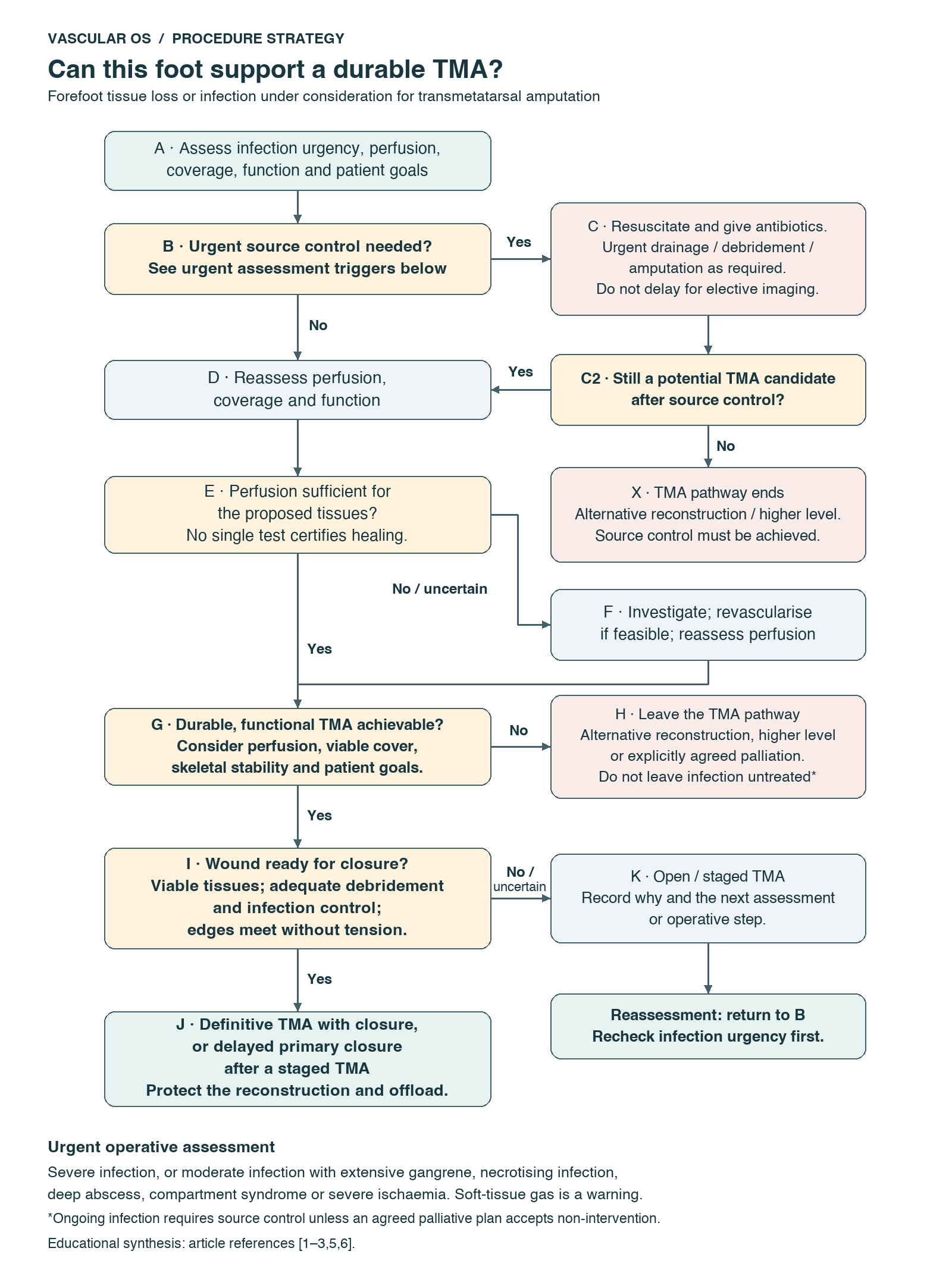
Strategy pathway: Vascular OS educational synthesis of [1–3,5,6]. Urgent source control may require a higher amputation. During staged management, reassess infection urgency as well as perfusion, coverage and function. Ongoing infection still requires source control unless an explicitly agreed palliative plan accepts non-intervention. No single perfusion result certifies healing.
Setup
- Confirm the side, planned level, revascularisation status and contingency for an open wound or higher amputation. Agree the anaesthetic approach and postoperative protection. Position supine. [3,4]
- Have a small amputation set, periosteal elevator, bone saw, bone rasp, haemostatic instruments and separate labelled specimen containers available. Prepare the whole foot and isolate grossly contaminated tissue where possible. [1,3,4]
- Distinguish perioperative antibiotic prophylaxis from treatment of established infection. In infection, start treatment promptly and obtain appropriate samples without delaying urgent care. [1,7]
- A tourniquet is selective: it can improve haemostasis but obscures assessment of tissue bleeding. If used, release it before judging final perfusion and haemostasis. [1,3,4]
Perform
Phase A — Mark and preserve the plantar flap
Mark a shorter dorsal and longer plantar component through viable tissue, allowing the plantar flap to cover the final bone ends without tension. Adapt the design to the actual defect; do not retain infected skin merely to complete a standard fish-mouth incision. [1,3,4]
Incise full thickness with minimal undermining. Handle the skin gently and preserve the plantar padding. A thin or crushed flap may close initially and then fail. [1,3]
Phase B — Expose and divide the metatarsals
Divide the dorsal tendons and expose the planned bone cuts with limited periosteal stripping. Protect the plantar tissues from the saw. Remove enough bone to clear disease and permit coverage while retaining useful length. [1,3,4]
Create a smooth metatarsal contour and avoid an isolated prominent stump. Published operative descriptions differ: one preserves a natural parabola with the second metatarsal longest, while another uses progressive lateral shortening. Do not apply a fixed millimetre pattern at the expense of disease clearance, viable coverage or pressure avoidance. [1–3]
Bevel the cuts from dorsal-distal to plantar-proximal: the plantar edge should finish more proximally than the dorsal edge. Smooth sharp edges and the medial first/lateral fifth metatarsal prominences. This reduces focal pressure against the flap. [1–3]
Phase C — Remove the forefoot and clear residual disease
Release the plantar attachments close to the bone, keeping the flap intact. Excise non-viable tissue and redundant poorly vascularised tendon/sheath without stripping useful padding. Inspect for infection tracking proximally beyond the visible forefoot lesion. [1–4]
When operating for infection, obtain an aseptic deep-tissue specimen when microbiology will guide therapy. If osteomyelitis is present or suspected, obtain appropriate bone specimens for culture. When retained-margin status will alter treatment, sample proximal retained bone aseptically and label it separately from removed infected bone; consider histology where clinically useful. A bleeding bone end does not by itself exclude residual osteomyelitis. [2–4,6]
Phase D — Check perfusion, haemostasis and mechanical balance
Irrigate, secure bleeding points and reassess tissue viability. Test flap coverage over the actual residual bones. If it is tight, revise the bone level or reconstruction; forcing the skin together is not an acceptable solution. [1,3,4]
Reassess ankle position. Consider Achilles lengthening or other balancing procedures when deformity or loading threatens durability. Observational TMA data, predominantly in people with diabetes, suggest fewer late forefoot ulcers with concomitant lengthening, but selection was non-random and no universal indication is established; weigh this against tendon rupture, heel ulceration and the additional protection required. It is not a mandatory component of every TMA. [2,3,8]
Phase E — Close only when the wound is ready
Close with interrupted sutures only when the remaining tissues are viable, infection has been adequately debrided and controlled, and the edges meet without tension. Aim for a well-padded stump with the scar away from the plantar loading surface. Drain use is selective and does not replace haemostasis. [1,3,4]
Leave the wound open if contamination, residual infection, uncertain viability or inadequate coverage makes closure unsafe. Record the reason and the next assessment or operative step. Apply a protective, non-constricting dressing; a suitably padded splint near neutral may help limit equinus when compatible with the wound. [1–4]
Pearls & Pitfalls
- Preserve useful length: retain bone only if it is healthy and can be covered by viable tissue. [1,3]
- Preserve flap thickness: excessive undermining and cosmetic trimming can compromise perfusion. [1,3]
- Check the plantar bone edge: a satisfactory dorsal appearance can conceal a plantar prominence. [1–3]
- Stage when necessary: an open wound with a clear reconstruction plan is preferable to closing over infection or under tension. [1–3]
- Plan for loading: healing of the incision does not eliminate the risk from neuropathy, deformity or poor footwear. [2,3,8]
Completion
Before leaving theatre, confirm and document: [1–4]
- Diseased tissue removed; retained bone and soft-tissue margins assessed and indicated specimens clearly labelled.
- Flap viability, haemostasis, smooth bone contour and absence of closure tension.
- Open versus closed management, any tendon procedure, dressing and splint position.
- Antibiotic plan, loading restrictions, wound-review timing and the trigger for return to theatre or further perfusion assessment.
Complications & Rescue
Bleeding or haematoma: assess haemodynamic impact and inspect the wound. A tense collection can compromise the flap; significant ongoing bleeding or an expanding haematoma requires prompt operative haemostasis and evacuation. [1,3]
Dusky flap or wound-edge necrosis: remove external constriction and assess tension, perfusion, haematoma and infection. Reassess new ischaemia or progressive tissue loss urgently with pedal Doppler signals, appropriate perfusion testing and arterial imaging. Do not repeatedly trim the wound without addressing its cause. [1–3,5]
Persistent infection or sepsis: reassess the whole foot for an undrained compartment or infected bone. Obtain deep specimens, revise antibiotics and perform further source control. A higher amputation may be necessary if the remaining foot cannot be cleared of infection or reconstructed. [1–3,6]
Non-healing or recurrent plantar ulceration: assess perfusion, infection, bone prominence, equinus and actual loading in the patient's footwear. Offload promptly; investigate suspected osteomyelitis and correct the mechanical cause where feasible. Antibiotics alone will not correct pressure-related breakdown. [1–3,6]
Postoperative Management
Protect the reconstruction
Specify the loading plan rather than writing only “mobilise”; physiotherapy should plan safe transfers and walking aids. For an uncomplicated, primarily closed TMA without an additional tendon procedure, a conservative planning guide is non-weight-bearing until the incision heals, often around 4–6 weeks. Earlier protected heel-only loading in an offloading shoe may be prescribed selectively. Progression depends on flap viability, perfusion and wound healing. [1,3,10,11]
Arrange an early wound review and earlier inspection for bleeding, deterioration or systemic illness. Protect both heels and the contralateral foot. Assess analgesia, glycaemic control, nutrition, renal function and venous thromboembolism risk; provide appropriate prophylaxis and PAD secondary prevention. [1,2,4]
Treat the infection that remains
Use the operative findings, available margin culture/histology and clinical response to determine treatment with microbiology. For diabetes-related osteomyelitis, IWGDF/IDSA distinguishes: [6]
| Residual infection after surgery | Antibiotic approach |
|---|---|
| Infected bone completely resected, without residual soft-tissue infection needing longer treatment | Short course, usually 2–5 days |
| Positive culture or histology at the retained bone margin | Consider up to 3 weeks |
| Unresected osteomyelitis or retained dead infected bone | Usually 6 weeks |
The evidence for postoperative bone-infection durations is limited. Interpret a positive margin in context, including possible contamination. Residual soft-tissue infection has its own treatment requirement. [6]
UK practice — differing duration frameworks: NICE NG19 Table 2 uses at least 7 days for moderate/severe diabetic foot infection and up to 6 weeks for osteomyelitis, guided by clinical assessment. It does not specify a separate course after complete bone resection; IWGDF/IDSA Table 5 gives 2–5 days in that setting. Agree and document duration with microbiology under local antimicrobial policy, using resection completeness, margin results, residual soft-tissue infection and clinical response. Review intravenous treatment by 48 hours. [6,7]
For a selected open postoperative wound, negative-pressure therapy may assist healing after debridement. It does not replace perfusion, source control or antibiotics for infection. [2,6,7]
Make healing durable
Once the wound allows, arrange orthotic footwear and an appropriate forefoot filler/pressure-relieving insole. Review fit, gait, ankle position and recurrent callus with the foot-care and rehabilitation team. Continue surveillance of both feet and teach daily skin inspection and prompt reporting of new wounds. [1–3]
Remember
- Choose the most distal level that can heal and function. [1–3]
- Urgent infection needs source control; definitive reconstruction also needs perfusion. [2,3,5]
- Preserve plantar padding and remove pressure-producing bone edges. [1–3]
- Close only viable tissue after adequate debridement and infection control, without tension. [1,3]
- Long-term success depends on loading, footwear and surveillance as well as wound closure. [1–3,8]
References
- Sidawy AN, Perler BA, editors. Rutherford's Vascular Surgery and Endovascular Therapy. 10th ed. Elsevier; 2023.
- Loftus I, Hinchliffe RJ, editors. Vascular and Endovascular Surgery: A Companion to Specialist Surgical Practice. 7th ed. Elsevier; 2024.
- Chaikof EL, Cambria RP, editors. Atlas of Vascular Surgery and Endovascular Therapy: Anatomy and Technique. Saunders; 2014.
- Forsyth J. How to Be a Safe Consultant Vascular Surgeon from Day One: The Unofficial Guide to Passing the FRCS (Vasc). 1st ed. CRC Press; 2023.
- IWGDF, ESVS, SVS. Intersocietal guideline on peripheral artery disease in people with diabetes mellitus and a foot ulcer. 2023. Official guideline.
- Senneville E, Albalawi Z, van Asten SA, et al. IWGDF/IDSA guidelines on the diagnosis and treatment of diabetes-related foot infections (2023). Clin Infect Dis. 2023:ciad527. doi:10.1093/cid/ciad527. Official IWGDF guideline PDF.
- National Institute for Health and Care Excellence. Diabetic foot problems: prevention and management. NG19. 2015; updated 2019. Recommendations.
- Bullock MJ, Gill CM, Thomas R, Blebea J. Concomitant Achilles tendon lengthening with transmetatarsal amputation for the prevention of late forefoot ulceration. J Bone Joint Surg Am. 2022;104:1722–1729. doi:10.2106/JBJS.21.00888.
- Coye T, Ansert E, Suludere MA, Chung J, Kang GE, Lavery LA. Healing rates and outcomes following closed transmetatarsal amputations: a systematic review and random effects meta-analysis of proportions. Wound Repair Regen. 2024;32:182–191. doi:10.1111/wrr.13143.
- Attinger CE, Evans KK. Operative management of diabetic foot wounds and infections: maximizing length and optimizing biomechanics. In: Sidawy AN, editor. Diabetic Foot: Lower Extremity Arterial Disease and Limb Salvage. 1st ed. Lippincott Williams & Wilkins; 2006.
- UW Health. Transmetatarsal Amputation (TMA) and Toe Amputation. Health Facts for You, no. 4892. Activity advice.