Diabetic Foot Ulcer with PAD
Diabetic foot ulceration: infection, ischaemia, offloading and limb preservation.
Written September 1, 2026 · Mr Razhan K Ali
At a glance
A diabetic foot ulcer reflects interacting neuropathy, mechanical loading, impaired perfusion and sometimes infection. Treat each contributor together: antibiotics cannot drain an abscess, angioplasty cannot remove plantar pressure, and a dressing cannot compensate for either. [1–6]
At first assessment establish stability, source-control need, perfusion, pressure and whether care is deliverable. Assess wound dimensions, infection grade, objective perfusion and offloading. [1–5,10]
- Act immediately for sepsis, necrotising infection, deep abscess, wet/infected gangrene or an acutely threatened limb. In UK practice, NICE also requires immediate acute-service assessment for any gangrene, ulceration with limb ischaemia, or concern for deep soft-tissue/bone infection. Involve the on-call vascular consultant when source control, perfusion treatment or the level of salvageable tissue requires an urgent operative decision. Do not await MRI before draining a surgical emergency. [3,4,10]
- Assess PAD in every ulcer, even when the foot is warm or the patient has no rest pain. Combine pedal Doppler waveforms, ankle measurements and toe measurements; a reassuring single test does not exclude disease. [1–3]
- Diagnose infection clinically. Colonisation or a positive swab alone does not justify antibiotics. A clinically uninfected ulcer needs wound care, pressure relief and perfusion assessment. [4]
- Offload immediately, adapting the method to location, infection, ischaemia and inspection needs. A non-removable knee-high device is first-line for an uncomplicated neuropathic plantar forefoot/midfoot ulcer; it is not a universal device for every diabetic wound. [5,10]
- Measure progress. Deterioration demands prompt reassessment. Less than 50% reduction in area after four weeks of appropriate care is a trigger to reconsider perfusion and the whole treatment plan, not a reason to wait four weeks before treating ischaemia. [3]
Understand
Why the foot ulcerates
Sensory neuropathy removes protective pain; deformity and joint stiffness concentrate loading; autonomic dysfunction promotes dry, fissured skin. Repetitive pressure produces callus and breakdown, sometimes concealing a deep cavity. Examine the shoe for a tight toe box, foreign body or seam, and check offloading devices for injury. [1,2]
PAD impairs tissue tolerance and healing. Calcified distal disease can falsely elevate ankle pressures; assess the tibial and pedal circulation. Neuropathy can mask rest pain, while infection or autonomic changes leave an ischaemic foot warm. Describe the foot as neuropathic, ischaemic or neuroischaemic, then grade infection separately. [1–3]
Infection can track along tendons and through compartments despite little erythema or fever. A chronic deep ulcer over bone, exposed bone or a swollen “sausage” toe suggests contiguous osteomyelitis. [1,2,4]
What to establish at the bedside
History: onset and progression; preceding trauma or new footwear; previous ulcers, amputations and revascularisation; discharge, erythema, systemic illness and recent antibiotics; mobility, usual walking and pressure exposure. Establish diabetes treatment, renal disease/dialysis, cardiovascular disease, smoking, nutritional concerns, allergies and previous resistant organisms. Ask what the patient can manage at home, including help with dressings, vision, balance, stairs and transport. These determine whether an apparently suitable outpatient plan is deliverable. [1–5,10]
Patient: assess observations, organ dysfunction, hydration, cognition, pain and glucose; investigate ketones/acidosis when indicated by acute illness or diabetes medicines. Usually obtain FBC, renal function/electrolytes and CRP, adding tests for sepsis or operative risk as needed. Normal inflammatory markers do not exclude foot infection or osteomyelitis. [1,2,4,10]
Both feet and both shoes: inspect plantar surfaces, heels, interdigital spaces and nail folds. Record ulcer site, length, width, depth, tissue, exudate, surrounding callus/maceration, undermining, exposed structures and gangrene. Photograph with consent and a scale, and measure consistently after appropriate cleansing/debridement. Look for spreading infection, fluctuance, crepitus, tendon involvement and deformity; assess protective sensation with a 10-g monofilament and an additional neurological test when appropriate. Palpate pulses, record Doppler signals and arrange objective pressures. [1–4,8,10]
Examine the mechanical cause: flexible versus fixed toe deformity, prominent metatarsal head, limited ankle dorsiflexion, restricted first metatarsophalangeal movement, Charcot collapse and altered loading after amputation. Record heel pressure from bed rest separately from pressure during walking. This examination should explain the offloading prescription. [1,2,5]
Describe severity without confusing classification systems
Use SINBAD for concise ulcer description and communication: site, ischaemia, neuropathy, bacterial infection, area and depth. Record its components, not just a total. NICE also accepts the University of Texas system and advises against Wagner for ulcer assessment. Neither replaces an infection grade or a vascular assessment. [9,10]
Use IWGDF/IDSA for infection. Infection is supported by at least two compatible local inflammatory findings—swelling/induration, erythema, tenderness/pain, warmth or purulent discharge—after considering alternatives such as trauma, gout and Charcot. For erythema to count in the mild-infection definition, it extends >0.5 cm but <2 cm from the wound margin; redness ≥2 cm is a moderate-infection feature. Confirmed osteomyelitis is classified as grade 3(O) or 4(O), even when the usual local inflammatory signs are absent. [4]
| Infection category | Practical definition | Consequence |
|---|---|---|
| Uninfected, grade 1 | No clinical infection | No antibiotics for the ulcer |
| Mild, grade 2 | Skin/subcutaneous tissue only, no systemic manifestations; if erythema is present, >0.5 cm but <2 cm from the wound margin | Usually oral treatment and close outpatient review if the rest of the assessment permits |
| Moderate, grade 3 | Erythema reaches ≥2 cm and/or infection extends deeper than skin/subcutaneous tissue; no systemic inflammatory response | Assess urgently for drainage, bone/joint involvement and PAD; admission often needed with PAD or other important morbidity |
| Severe, grade 4 | Foot infection with at least two SIRS criteria | Admit and treat urgently; assess organ dysfunction and sepsis as well |
The SIRS criteria used by this classification are temperature >38°C or <36°C, heart rate >90/min, respiratory rate >20/min or PaCO₂ <32 mmHg, white count >12 or <4 ×10⁹/L or >10% immature forms. This is an infection-grading definition, not a substitute for contemporary sepsis assessment: a patient with dangerous organ dysfunction needs urgent treatment regardless of whether two SIRS criteria are recorded. Add “(O)” to grade 3 or 4 when osteomyelitis is present. [4]
For PAD with tissue loss, use WIfI: Wound, Ischaemia and foot Infection. It supports limb-threat staging and discussion of revascularisation benefit. Its infection component runs from 0 to 3, whereas IWGDF/IDSA runs from 1 to 4. Use the published WIfI matrix to derive clinical stage 1–4; do not simply add the three numbers. [3,9]
| WIfI component | Useful bedside anchors |
|---|---|
| Wound | Grade 1: small, shallow wound, no gangrene; grade 2: deeper tissue loss, shallow heel ulcer or gangrene confined to digits; grade 3: extensive forefoot/midfoot loss or full-thickness heel injury, potentially involving calcaneus |
| Ischaemia | Toe pressure ≥60, 40–59, 30–39 and <30 mmHg correspond to grades 0, 1, 2 and 3 respectively; use the full table when other tests are needed |
| Foot infection | None, mild, moderate, severe → grades 0, 1, 2, 3 |
WIfI informs judgement; it does not predict an individual patient's outcome with certainty. [3,9]
Decide
Use the pathways with the decision rules below; reassess whenever the clinical picture changes.
Algorithm 1 — the first encounter
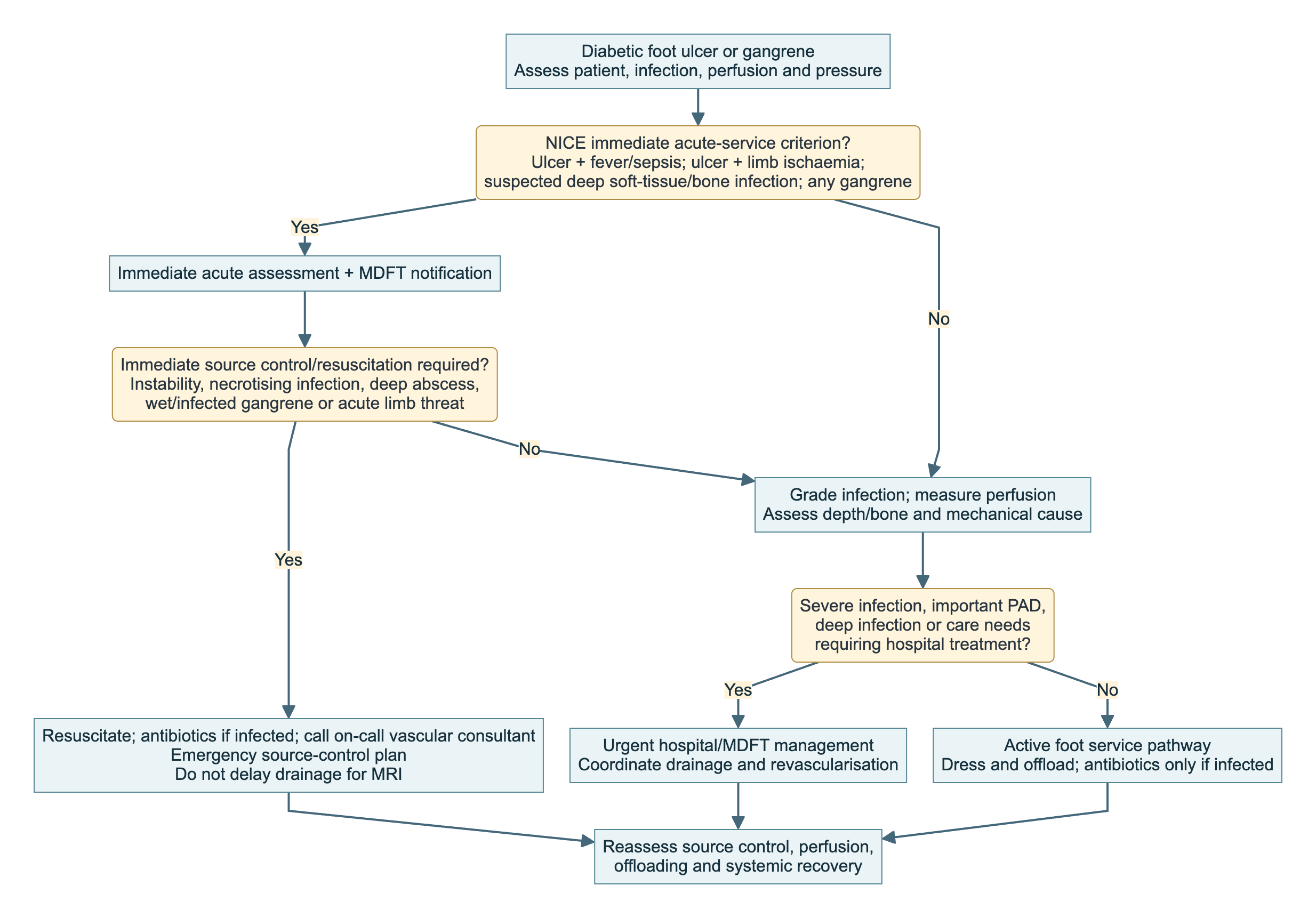
In words: separate referral urgency from operative urgency. NICE requires immediate acute assessment and multidisciplinary foot care team (MDFT) notification for ulceration with fever/sepsis or limb ischaemia, suspected deep soft-tissue/bone infection, or any gangrene. A stable dry gangrenous toe does not automatically need immediate debridement. Admit severe infection and consider admission for moderate infection with PAD or important morbidity. Osteomyelitis alone does not invariably require admission or intravenous therapy. [4,10]
Other active diabetic foot problems should enter the specialist pathway within one working day, with triage within a further working day. For an inpatient, involve the multidisciplinary foot service within 24 hours. These are access standards, not permission to defer treatment of an emergency. [10]
Algorithm 2 — is perfusion sufficient, and when should I revascularise?
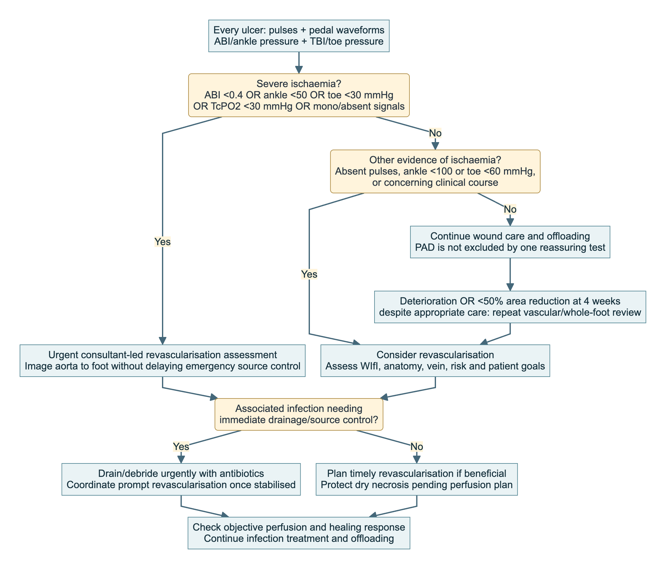
An ABI of 0.9–1.3, TBI ≥0.70 and biphasic/triphasic pedal signals make PAD less likely; they do not rule it out. Calcification can make ankle pressures falsely reassuring. Where toe pressure is unavailable or unsuitable, use TcPO₂ or skin perfusion pressure alongside the clinical assessment. [3]
Act on ischaemia: consider revascularisation with absent pulses, ankle pressure <100 mmHg or toe pressure <60 mmHg. Urgent consultant-led assessment is indicated for ABI <0.4, ankle pressure <50, toe pressure <30 or TcPO₂ <30 mmHg, or monophasic/absent pedal signals. Interpret these with the clinical course. [3]
Keep prognostic and action thresholds separate. Toe pressure ≥30 mmHg, TcPO₂ ≥25 mmHg and skin perfusion pressure ≥40 mmHg support healing probability; none guarantees healing. The dedicated PAD guideline nevertheless uses TcPO₂ <30 mmHg among its severe-ischaemia triggers for urgent assessment. A value of 27 is therefore not reassuring simply because it exceeds the prognostic value of 25. [3]
Plan revascularisation from inflow to the pedal circulation, identifying usable targets. Select duplex, CTA, MRA or catheter angiography around calcification, renal function and the detail needed; obtain adequate pedal imaging. Weigh operative risk, vein conduit, WIfI, anatomy and functional goals together. [3]
Sequence depends on the threat. Drain a deep abscess, necrotising infection or wet gangrene urgently, then coordinate prompt revascularisation once stabilised, often within days. Protect stable, dry, uninfected ischaemic eschar while defining perfusion and tissue strategy: aggressive debridement may create a nonhealing wound. New infection changes that balance immediately. [1–4]
Algorithm 3 — suspected osteomyelitis
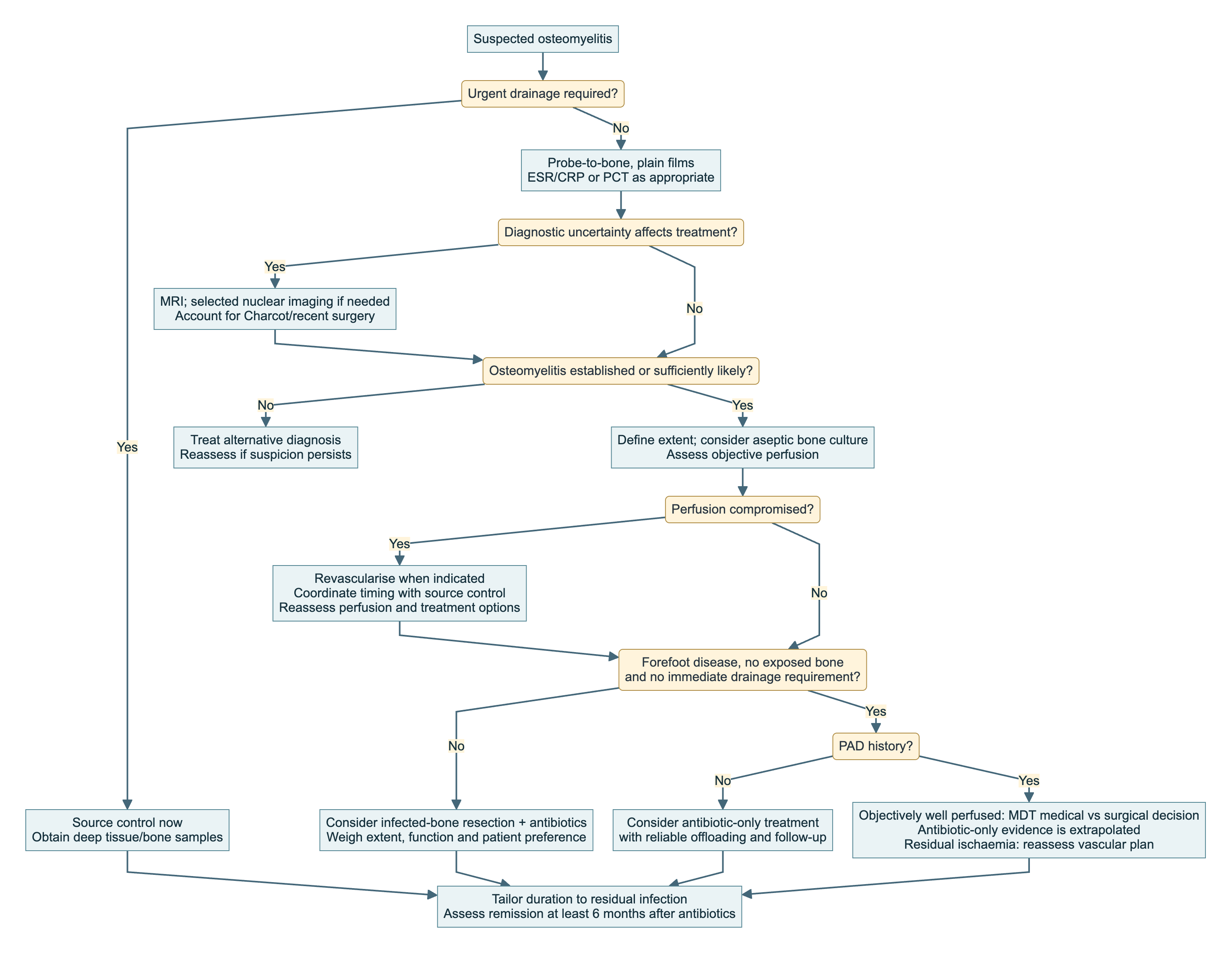
Probe-to-bone changes probability: a positive result supports osteomyelitis in a high-risk wound, but a negative result cannot exclude it when suspicion remains high. Plain films assess bone, deformity, foreign material and gas; early films may be normal. Use MRI when diagnostic uncertainty affects treatment, recognising reduced specificity with Charcot or recent surgery. [4,10]
Obtain bone aseptically at surgery or percutaneously when appropriate for culture, with histology in selected cases. A superficial swab cannot identify bone pathogens reliably. Agree sampling with microbiology; no antibiotic-free interval is universally established, and urgent treatment must not be withheld to improve yield. [4]
Consider antibiotic-only treatment for forefoot osteomyelitis without PAD, exposed bone or an immediate drainage requirement. These are the formal recommendation's selection criteria. When perfusion is compromised, revascularise if indicated and reassess objectively. A history of PAD does not mandate bone resection in a now well-perfused foot, but antibiotic-only treatment here extrapolates beyond the trial population and requires explicit MDT judgement. [3,4]
Algorithm 4 — choose the offloading method
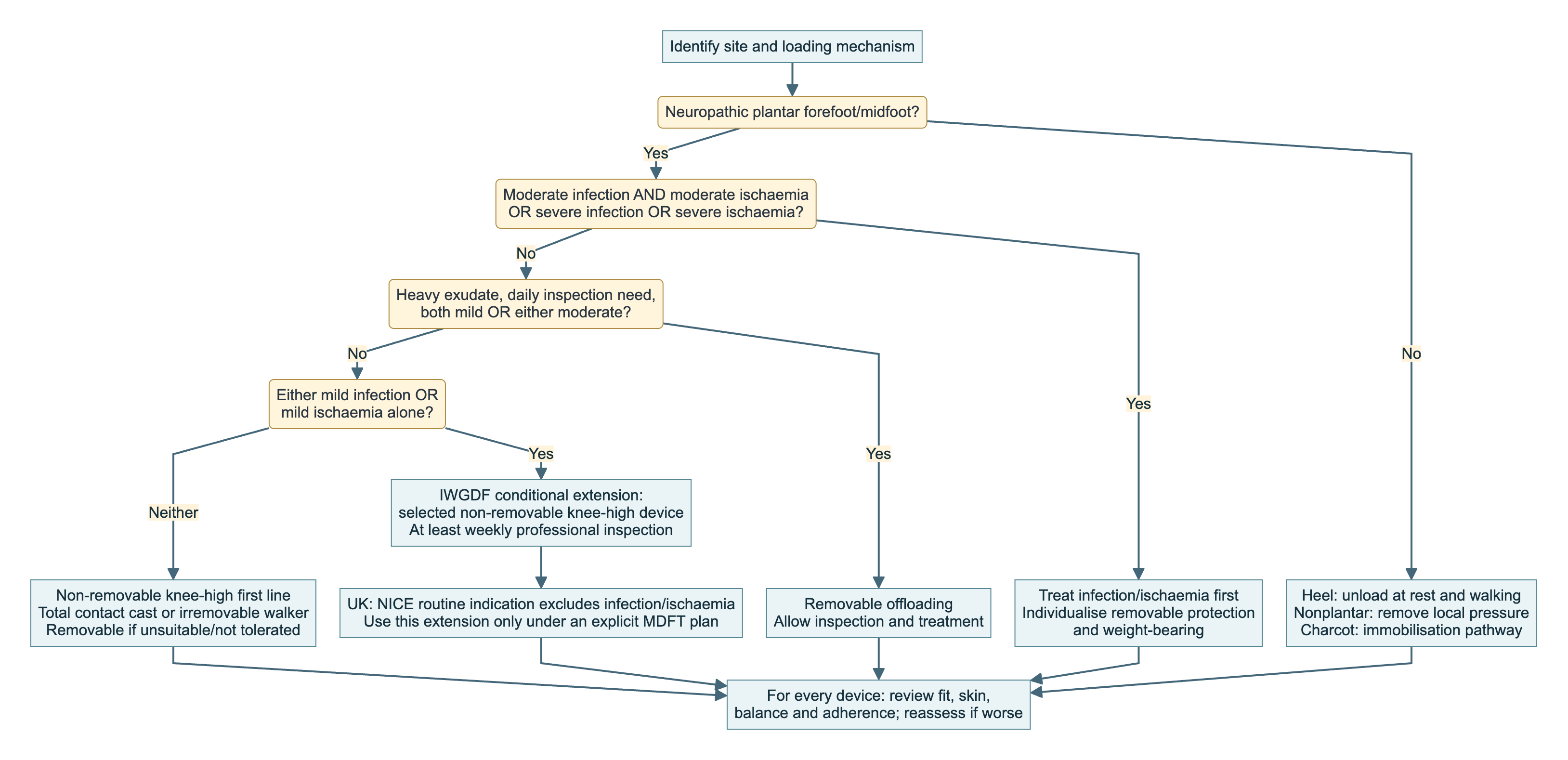
Non-removable devices improve adherence. Choose a total contact cast or walker made non-removable according to expertise, deformity and circumstances. If unsuitable or intolerable, use a removable knee-high or ankle-high device for every weight-bearing activity, including short indoor journeys. If devices are unavailable, suitable footwear plus felted foam is a lower-order option; felt alone is not equivalent. [5]
IWGDF conditionally permits a non-removable knee-high device where there is either mild infection or mild ischaemia alone, with at least weekly professional review. Both mild infection and mild ischaemia together, either moderate condition, heavy exudate or frequent inspection requirements favour removable offloading; both moderate conditions together, or either severe condition, require infection/ischaemia treatment first with individualised removable protection. The “mild/moderate” ischaemia modifiers require clinical judgement and should not be mechanically replaced by a WIfI number. [5]
UK divergence: NICE's routine non-removable-cast indication is narrower—neuropathic, uninfected and non-ischaemic plantar forefoot/midfoot ulceration. Any extension beyond this should be an explicit MDFT plan where inspection and rapid reassessment are reliable. [5,10]
Check falls risk, contralateral disease and limb-length discrepancy; consider walking aids and a contralateral shoe lift. Review device fit, edges and both feet, and provide access to urgent device removal/reassessment if deterioration occurs. [5]
Algorithm 5 — when pressure-relieving surgery is appropriate
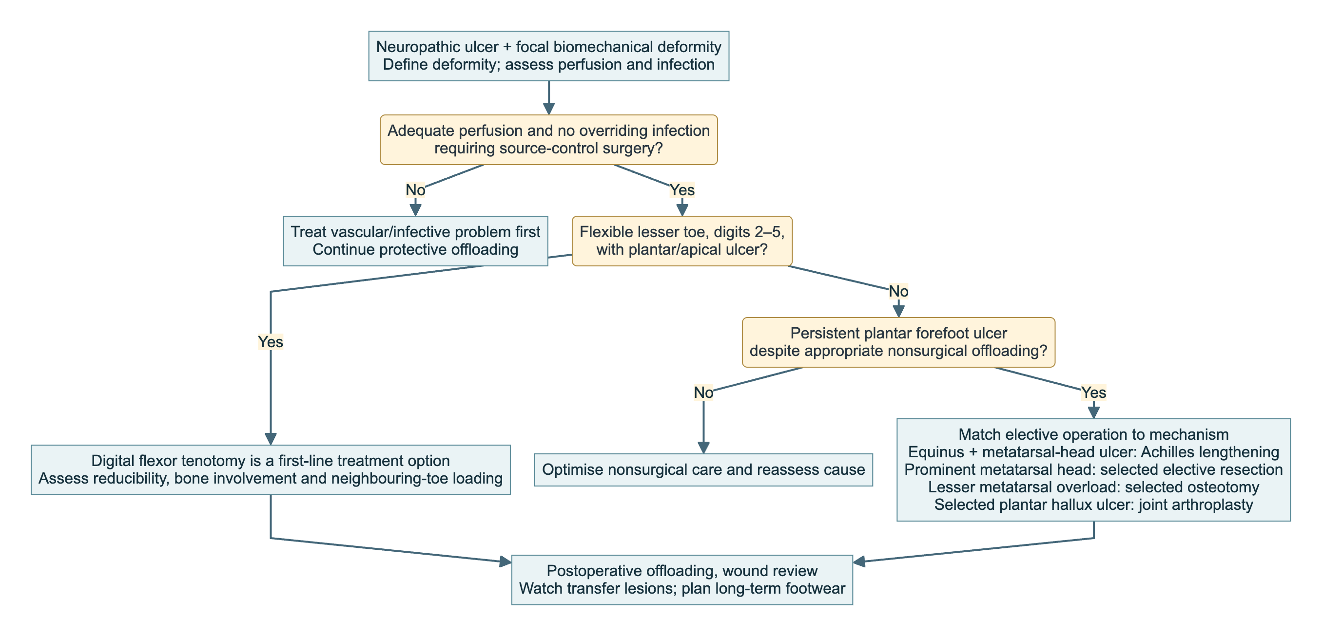
Digital flexor tenotomy: a first-line option for a neuropathic plantar/apical ulcer on a flexible lesser toe (digits 2–5), with strong recommendation/moderate-certainty evidence. Prior failure of nonsurgical treatment is not required. Do not extrapolate to the hallux, rigid clawing or a toe needing infection-control surgery. Check reducibility, perfusion and bone involvement; discuss transfer lesions and maintain footwear/surveillance. Preventive tenotomy for selected pre-ulcerative lesions is a separate conditional recommendation. [1,5,8]
Achilles tendon lengthening: consider with continued offloading for a neuropathic plantar metatarsal-head ulcer after appropriate nonsurgical offloading fails, particularly with restricted ankle dorsiflexion. It reduces forefoot loading; it does not treat a plantar heel ulcer. The recommendation is conditional/moderate certainty. Balance benefit against weakness, altered gait and heel transfer ulceration; avoid overlengthening. Confirm adequate perfusion, infection control and ability to complete postoperative protection and rehabilitation. [1,5]
Other operations: match metatarsal-head resection, lesser-metatarsal osteotomy or hallux joint arthroplasty to the pressure mechanism after nonsurgical offloading fails. Continue postoperative offloading and monitor transfer lesions. Resection for osteomyelitis/septic joint is an infection-control indication, distinct from elective offloading. Severe ischaemia contraindicates elective surgical offloading. [1,5]
Justify
Which guidelines govern this article?
The 2023 PAD guideline is jointly IWGDF/ESVS/SVS: it is one intersocietal pathway, not competing ESVS and IWGDF advice. It governs vascular assessment, healing prognosis and revascularisation. Other IWGDF documents cover offloading, wound healing, prevention, classification and Charcot; infection guidance is joint IWGDF/IDSA. NICE NG19 adds UK standards and prescribing guidance. [3–10]
| Source | Decision it answers | Important qualification |
|---|---|---|
| Joint IWGDF/ESVS/SVS PAD 2023 | Is ischaemia contributing; how urgently to investigate; whether/how to revascularise | Bedside thresholds change probability or urgency; none guarantees healing |
| IWGDF/IDSA infection 2023 | Is it infected; how severe; sampling, antibiotics, drainage and bone infection | Antibiotics alone cannot provide source control or restore perfusion |
| IWGDF offloading 2023 | Which device or corrective operation addresses the ulcer's loading mechanism | Recommendation varies with site, infection, ischaemia and ability to inspect |
| IWGDF wound healing 2023 | Which adjuncts may help after best standard care | Most adjunct recommendations are conditional; correct the cause of failure first |
| IWGDF Charcot/prevention/classification 2023 | Immobilisation, remission, recurrence prevention and shared description | Charcot guideline recommendations principally concern intact skin |
| NICE NG19 | UK urgency, organisation, ulcer care and antimicrobial choices | Some adjunct and casting recommendations differ from IWGDF |
A strong recommendation means confidence that the balance of benefits and harms favours the recommended course; it does not necessarily mean high-certainty evidence. Conditional recommendations require closer attention to selection, preferences and resources. Best-practice statements support several urgent decisions where trials are impractical. [3–8]
Differences that change practice
Perfusion thresholds: use the dedicated PAD guideline for exact vascular action thresholds. It distinguishes TcPO₂ ≥25 mmHg as favourable prognostic information from <30 mmHg as a severe-ischaemia trigger. Its four-week healing reassessment also takes precedence here over the broader practical summary's four-to-six-week wording. [3]
Casting: IWGDF permits selected non-removable offloading with either mild infection or mild ischaemia alone, with close monitoring; NICE's routine indication excludes both. Do not use that conditional extension where the wound needs frequent access or the clinical trajectory is uncertain. [5,10]
Adjuncts: IWGDF conditionally supports selected hyperbaric oxygen after standard care fails in ischaemic/neuroischaemic ulcers; NICE restricts it to trials. NICE also restricts platelet-rich plasma gel, regenerative wound matrices and growth factors to trials, while allowing selected dermal/skin substitutes for stalled healing on MDFT advice. IWGDF's leucocyte/platelet/fibrin patch and placental-product recommendations concern specific interventions: do not assume product categories are interchangeable. Check exact product guidance and local governance. The July 2025 NICE decision to update topical-oxygen guidance is not itself a treatment recommendation. [6,10]
Bypass or endovascular treatment?
The joint PAD guideline conditionally favours bypass when infrainguinal revascularisation is indicated, a suitable single-segment saphenous vein is available and the patient is suitable for either strategy. This is not a universal bypass-first rule. Patient risk and preference, limb threat, anatomy, conduit and local ability to provide both approaches remain central. [3]
In BEST-CLI, the suitable-vein cohort had fewer major adverse limb events or deaths with surgery than endovascular treatment: 42.6% versus 57.4%, hazard ratio 0.68. The composite included major limb reinterventions; this is not simply an amputation-free-survival comparison. In cohort 2, without adequate single-segment GSV, the primary outcome occurred in 42.8% versus 47.7%, HR 0.79 (95% CI 0.58–1.06); failure to detect a statistically significant difference does not establish equivalence. [11]
In BASIL-2, 345 patients requiring infrapopliteal revascularisation had more major amputations or deaths with vein-bypass-first than endovascular-first: 63% versus 53%, adjusted hazard ratio 1.35; the difference was largely driven by mortality. Recruitment limitations add uncertainty. These CLTI trials had different populations and endpoints, and neither consisted exclusively of diabetic ulcers. Use them to inform selection rather than declaring one technique universally superior. [11,12]
Restore inline flow to at least one foot artery. Targeting the ulcer's angiosome is conditional/very-low-certainty advice: consider the pedal arch, collaterals, target quality and durability. Do not compromise a sound reconstruction to pursue an angiosome or require all three tibial arteries to be opened. Confirm objective perfusion and wound progress afterwards. [3]
What newer evidence does not yet settle
Guidelines prefer an aseptically collected deep tissue specimen over a superficial swab. CODIFI2, published in 2025, recruited only 149 of its planned 730 participants and did not demonstrate a healing advantage for tissue sampling. It was underpowered and does not establish equivalence. Retain appropriate tissue sampling, but do not tell patients that its superiority in healing outcomes is proven. [4,13]
Perform
Establish source control and obtain useful samples
For an infected wound, cleanse and, where appropriate, debride before obtaining deep tissue by curettage or biopsy. NICE permits a deep swab when tissue cannot be obtained. Collect blood cultures when systemic infection warrants them, without delaying urgent antibiotics. Tell the laboratory the site, depth, recent antibiotics and whether a specimen is bone; a surface culture cannot reliably define the organisms in infected bone. [4,10]
Decide what must be drained or removed now and what can be reconstructed later. Explore the anatomical extent of infection, remove non-viable infected tissue and obtain representative samples. Extensive destruction may require a toe/ray, partial-foot or major amputation to control infection. Early surgery within 24–48 hours is supported for selected moderate/severe infections, but this is not a waiting interval for necrotising infection or a deep abscess. Reassess for residual infection and further debridement as the foot declares itself. [1,2,4]
When resecting infected bone, document what remains and obtain appropriately labelled margin specimens when indicated under the local surgical/microbiology protocol. The eventual antibiotic duration depends on residual infection, not simply on whether “an amputation” occurred. Preserve viable tissue where possible, while avoiding a residual foot that cannot heal, bear load or be accommodated in footwear. [1,4,14]
Prescribe antibiotics for the infection actually present
Base initial treatment on severity, previous cultures and antibiotics, likely organisms, renal function, allergy and local microbiology policy. Then narrow to useful culture results and clinical response. Mild, previously untreated infection in the UK usually requires streptococcal and staphylococcal coverage; NICE lists oral flucloxacillin 500 mg–1 g four times daily for seven days, with review and extension when clinically indicated. Confirm suitability and dosing for the individual patient rather than copying a regimen into every case. [4,10]
Moderate/severe disease, necrosis, prior antibiotic exposure or deep infection may require broader Gram-positive, Gram-negative and anaerobic cover; use the local diabetic-foot/sepsis regimen and microbiology advice. Add MRSA coverage when risk or microbiology warrants it. Do not routinely add empirical antipseudomonal therapy merely because the patient has diabetes: prior isolates, geography/exposure and local resistance matter. Severe illness usually starts with intravenous treatment; review the route at about 48 hours and switch to an active oral option when the patient and infection permit. [4,10]
| Clinical situation | Guideline duration framework |
|---|---|
| Soft-tissue infection | Recommendation 12: 1–2 weeks. Final Table 5 lists 2–4 weeks for moderate/severe infection; individualise to source control and response. NICE starts many regimens at 7 days with review |
| Adequately debrided moderate/severe soft-tissue infection | Approximately 10 days may suffice; this is a conditional suggestion based on limited evidence |
| Extensive but improving, slowly resolving infection or severe PAD | Selected extension to 3–4 weeks; reassess the reason for slow response |
| Infected bone completely removed, no residual soft-tissue infection | Short postoperative course, generally 2–5 days in the guideline table |
| Positive bone margin after minor amputation/resection | Up to approximately 3 weeks |
| Osteomyelitis with infected bone retained/no bone resection | Usually 6 weeks |
The final IWGDF/IDSA publication contains this duration difference between Recommendation 12 and Table 5; agree the course with microbiology according to severity, source control and response. Residual soft-tissue infection needs treatment even after complete bone removal. Do not continue antibiotics simply because the ulcer remains open. Reassess deterioration immediately, or failure to begin improving within 1–2 days, for undrained infection, ischaemia, unsuitable cover or an alternative diagnosis; avoid serial empirical extensions. [4,10]
Create a wound environment that can heal
Remove callus and devitalised tissue by skilled sharp debridement when appropriate, taking perfusion and pain into account. The frequency follows clinical need. Keep a stable, dry ischaemic eschar protected pending the vascular plan unless infection requires source control. Select a basic dressing that manages exudate, protects surrounding skin and maintains an appropriate moist environment in a healable wound; avoid maceration and repeated dressing trauma. [1–3,6]
Do not use topical antiseptic/antimicrobial dressings (including silver) or honey to promote ulcer healing: IWGDF recommendations are strong against these uses. NPWT may support postsurgical wound healing (conditional), but do not use it to promote healing of nonsurgical diabetic ulcers (strong). Neither NPWT nor local antimicrobials replaces source control or systemic infection treatment. [4,6]
After adequate standard care fails, consider only a suitable adjunct: sucrose-octasulfate for a noninfected neuroischaemic ulcer with insufficient improvement after at least two weeks, or selected topical oxygen, leucocyte/platelet/fibrin patches or placental products. These are conditional options, subject to the UK distinctions above. None replaces perfusion, source control or offloading. [6,10]
Address the whole patient
Address glucose management, nutrition, renal function, oedema, pain and smoking cessation. Acute illness and procedures require an explicit diabetes medicine plan. The PAD guideline advises against starting an SGLT2 inhibitor during active ulceration/gangrene and suggests considering temporary interruption in existing users until healing; discuss this with the diabetes team in the context of acute-illness precautions and cardiorenal benefit. Do not interpret it as withdrawal of all long-term cardiovascular diabetes therapy. [1–3]
For symptomatic PAD, prescribe vascular secondary prevention: appropriate antiplatelet therapy, lipid lowering, blood-pressure treatment and smoking support. Selected patients may benefit from low-dose rivaroxaban with aspirin when bleeding risk permits; reconcile this with any post-revascularisation regimen rather than inadvertently creating prolonged multiple-antithrombotic therapy. Glycaemic and cardiovascular targets should be individualised for frailty, renal disease and hypoglycaemia risk. [3]
Manage
Particular presentations that change the plan
Heel ulcer: determine whether loading occurs in bed, during walking or both. A forefoot offloading prescription does not necessarily unload the heel. Assess calcaneal involvement, heel-pad viability and perfusion early. Selected plantar rearfoot ulcers may benefit from a non-removable knee-high device, but the evidence is very low certainty; other wounds require a removable device or heel-suspension strategy that permits inspection. Calcaneal osteomyelitis and extensive heel loss may need specialist reconstruction, partial calcanectomy or amputation, judged against perfusion, functional potential and treatment burden. [1,2,4,5]
Hot swollen neuropathic foot: consider active Charcot, including when pain is minimal. Start knee-high immobilisation/offloading while arranging investigation; NICE advises non-weight-bearing until the specialist team starts definitive treatment. Arrange aids and assess whether the patient can manage this safely. Obtain appropriate foot/ankle radiographs, with weight-bearing views when safe, and MRI if suspicion persists despite normal films. Normal inflammatory markers do not exclude Charcot. When an ulcer is present, infection and Charcot may coexist; investigate both and adapt the device to inspection needs. [4,7,10]
The strongest Charcot device recommendations concern intact skin. Assess remission from inflammation, temperature trend and imaging together. NICE describes a sustained difference <2°C with no further radiographic progression as suggesting resolution; IWGDF does not endorse a single temperature cut-off. Resume loading gradually with footwear/orthoses. Consider specialist reconstruction for instability or deformity that cannot be protected. For active Charcot, IWGDF strongly recommends against alendronate, pamidronate, zoledronate, calcitonin, PTH and methylprednisolone; its recommendation against denosumab is conditional. NICE limits bisphosphonate treatment to trials. [7,10]
Dry gangrenous toe: NICE requires immediate acute assessment/MDFT notification for any gangrene. After assessment, protect genuinely dry, uninfected tissue while defining perfusion and the tissue plan. Select conservative care or planned minor amputation around healing potential, pain, function and preference. Wet/infected conversion requires urgent source-control reassessment. [1–4,10,14]
Atypical or unexplained ulcer: reconsider an unusual site, edge, pain or failure despite adequate perfusion, pressure relief and infection treatment. Malignancy or inflammatory disease may require biopsy/specialist assessment. Diabetes does not explain every wound. [1]
A foot that cannot be usefully salvaged: discuss major amputation or symptom-focused care when uncontrolled infection, unreconstructable ischaemia, tissue destruction, severe frailty or treatment burden makes a healed functional foot unrealistic. Consider whether limited revascularisation could improve healing at the proposed amputation level. The decision should include the patient's goals, rehabilitation potential, contralateral limb and the consequences of repeated unsuccessful procedures. [3,14]
Algorithm 6 — reassessment, failure and remission
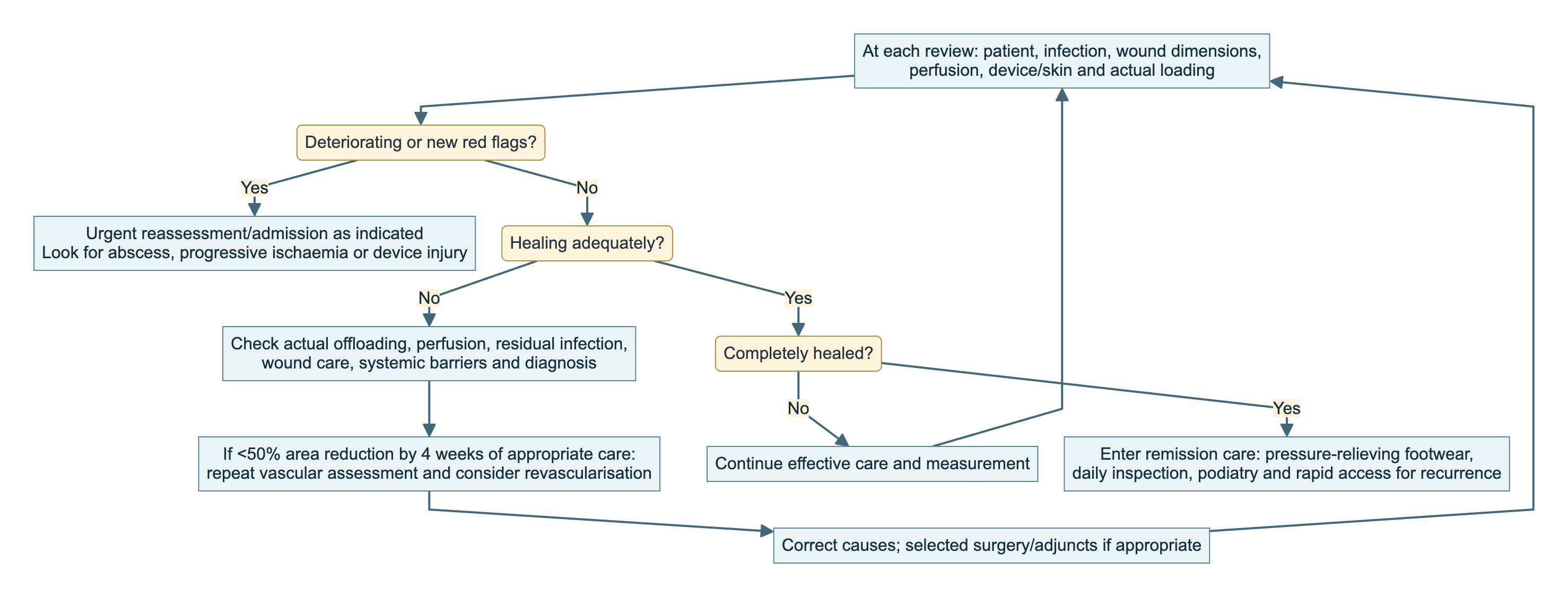
At every review assess the patient, infection, perfusion, wound and actual loading. Deterioration needs urgent reassessment; stalled healing requires correction of vascular, mechanical, infective and systemic barriers. If area falls by <50% after four weeks of appropriate care, repeat vascular assessment and reconsider revascularisation. Continue effective care until healed, then move to recurrence prevention. [1,3–6,8]
Measure area consistently: (baseline − current area) ÷ baseline area ×100. New undermining, necrosis or deeper infection overrides an apparently smaller surface area. An open artery with a nonhealing wound requires review of both perfusion and other healing barriers. [1,3]
Ask how many steps are taken without the prescribed device, whether the dressing becomes saturated, and whether the patient can attend follow-up. Treatment “failure” may reflect an impractical prescription. Review antimicrobial response early and active wounds according to severity, exudate and device needs; prevention-screening intervals are not appropriate intervals for an active infected ulcer. [4,5,8,10]
Give explicit return instructions for systemic illness, spreading redness/swelling, new discharge or odour with clinical deterioration, tissue darkening, device injury or a sudden change in foot appearance. Neuropathy means that absence of pain is not reassurance. [1,2,4,10]
After healing: remission is an active phase of care
Arrange footwear and insoles that reduce pressure at the previous ulcer site, ideally with demonstrated plantar-pressure reduction for a healed plantar ulcer. They must be worn indoors as well as outside. Review deformity and callus, treat pre-ulcerative lesions, and reinforce daily inspection of both feet and footwear; involve a carer when vision or mobility prevents self-care. Avoid barefoot walking and self-treatment of callus with blades or caustic agents. [8]
The IWGDF prevention schedule is annual review for risk 0, every 6–12 months for risk 1, every 3–6 months for risk 2, and every 1–3 months for risk 3. A previous ulcer in a patient with neuropathy or PAD places them in the highest-risk group. NICE preventive review intervals are annual for low risk, every 3–6 months for moderate risk, every 1–2 months for high risk without immediate concern, and every 1–2 weeks for high risk where there is immediate concern. These are preventive surveillance intervals, not appropriate review intervals for an active ulcer. Recurrent breakdown needs reassessment of its mechanism, not just another dressing. [8,10]
After treated osteomyelitis, assess remission over at least six months after antibiotic completion. A healed surface alone is not the entire follow-up assessment; monitor for recurrent drainage, swelling and new breakdown, while maintaining pressure relief and vascular surveillance as indicated. [4]
Remember
- Describe infection, perfusion and pressure separately, then treat them together. [1–5]
- A painless, warm foot can still be ischaemic or deeply infected. [1–4]
- Drain the urgent infection and coordinate prompt restoration of perfusion. In UK practice, any gangrene requires immediate acute assessment; protect stable dry necrosis after assessment while the tissue plan is defined. [1–4,10]
- No single pressure excludes PAD or guarantees healing. Deterioration overrides scheduled reassessment. [3]
- Antibiotic-only osteomyelitis treatment has narrow selection criteria; antibiotic duration follows residual infection, not ulcer closure. [4]
- Match offloading and corrective surgery to the anatomical mechanism. Flexible lesser-toe tenotomy is a first-line option for the appropriate active ulcer; other elective surgical-offloading procedures generally follow failure of nonsurgical offloading. [5]
- Healing is the start of recurrence prevention. The end point is a functional, protected foot and a care plan the patient can sustain. [3,8,14]
References
- Sidawy AN, Perler BA, eds. Rutherford's Vascular Surgery and Endovascular Therapy. 10th ed. Elsevier; 2023. Chapters 116–118.
- Loftus I, Hinchliffe R, eds. Vascular and Endovascular Surgery: A Companion to Specialist Surgical Practice. 7th ed. Elsevier; 2024. Chapter 5.
- Fitridge R, Chuter V, Mills J, et al. The intersocietal IWGDF, ESVS, SVS guidelines on peripheral artery disease in people with diabetes and a foot ulcer. Diabetes Metab Res Rev. 2024;40(3):e3686. doi:10.1002/dmrr.3686. 2023 guideline PDF.
- Senneville E, Albalawi Z, van Asten SA, et al. IWGDF/IDSA guidelines on the diagnosis and treatment of diabetes-related foot infections (2023). Clin Infect Dis. 2023:ciad527. Final publication, including Table 5. IDSA recommendations.
- Bus SA, Armstrong DG, Crews RT, et al. Guidelines on offloading foot ulcers in persons with diabetes (IWGDF 2023 update). Diabetes Metab Res Rev. 2024;40(3):e3647. doi:10.1002/dmrr.3647.
- Chen P, Campillo Vilorio N, Dhatariya K, et al. Guidelines on interventions to enhance healing of foot ulcers in people with diabetes (IWGDF 2023 update). Diabetes Metab Res Rev. 2024;40(3):e3644. doi:10.1002/dmrr.3644.
- Wukich DK, Schaper NC, Gooday C, et al. Guidelines on the diagnosis and treatment of active Charcot neuro-osteoarthropathy in persons with diabetes mellitus (IWGDF 2023). Diabetes Metab Res Rev. 2024;40(3):e3646. doi:10.1002/dmrr.3646.
- Bus SA, Sacco ICN, Monteiro-Soares M, et al. Guidelines on the prevention of foot ulcers in persons with diabetes (IWGDF 2023 update). Diabetes Metab Res Rev. 2024;40(3):e3651. doi:10.1002/dmrr.3651.
- Monteiro-Soares M, Hamilton EJ, Russell DA, et al. Guidelines on the classification of foot ulcers in people with diabetes (IWGDF 2023 update). Diabetes Metab Res Rev. 2024;40(3):e3648. doi:10.1002/dmrr.3648.
- NICE. Diabetic foot problems: prevention and management (NG19). Recommendations. July 2025 surveillance decision on topical oxygen.
- Farber A, et al. Surgery or Endovascular Therapy for Chronic Limb-Threatening Ischemia. N Engl J Med. 2022;387:2305–2316. doi:10.1056/NEJMoa2207899.
- Bradbury AW, Moakes CA, Popplewell M, et al. A vein bypass first versus a best endovascular treatment first revascularisation strategy for patients with chronic limb threatening ischaemia who required an infra-popliteal, with or without an additional more proximal infra-inguinal revascularisation procedure to restore limb perfusion (BASIL-2): an open-label, randomised, multicentre, phase 3 trial. Lancet. 2023;401:1798–1809. doi:10.1016/S0140-6736(23)00462-2.
- Nelson EA, Brown ST, Everett CC, et al. CODIFI2: Randomised controlled trial to compare clinical and cost-effectiveness of swabs versus tissue sampling to inform management of infected diabetic foot ulcers. Diabet Med. 2025;42(5):e70009. doi:10.1111/dme.70009.
- Chaikof EL, Cambria RP. Atlas of Vascular Surgery and Endovascular Therapy. Elsevier; 2014. Chapter 52.